2016 Work
Illustrations for Comprehensive Management of Swallowing Disorders, 2nd edition, Chapter 30, Zenker And Other Esophageal Diverticula; Rebecca Howell, MD and John Paul Giliberto, MD
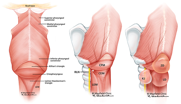
L-R: 1) Dorsal view of the hypopharynx. The apex of Killian’s triangle is formed between the oblique muscle fibers of the inferior pharyngeal constrictors and above the transversely oriented muscle fibers of the cricopharyngeus. Laimer-Haeckerman’s is an inverted triangle with the base immediately inferior to the transversely oriented cricopharyngeus and the apex directed inferiorly at a dehiscence between the longitudinal esophageal muscle (LEM). Printed with permission from Mayfield Clinic.
2,3) Posterolateral view of the hypopharynx. Killian-Jamison (KJ) is a lateral diverticulum that originates below the cricopharyngeus muscle (CPM) in a muscular gap (*) bounded inferiorly by the circular esophageal muscle (CEM) and medially by the longitudinal esophageal muscle (LEM). It is in this location that the recurrent laryngeal nerve (RLN) enters as it travels to the larynx. Zenker’s diverticulum is located in the posterior midline superior to the CPM as opposed to the Laimer’s diverticulum located inferior to the CPM. Printed with permission from Mayfield Clinic.
Infographic for The Pathophysiology of Delayed Cerebral Ischemia, Brandon Foreman, MD; published by The American Clinical Neurophysiology Society
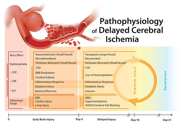
The Pathophysiology of Delayed Cerebral Ischemia CBF = Cerebral Blood Flow; CSD = Cortical Spreading Depolarization; CPP = Cerebral Perfusion Pressure; ICP = Intracranial Pressure; SIADH = Syndrome of Inappropriate Antidiuretic Hormone; SIRS = Systemic Inflammatory Response Syndrome Printed with permission from Mayfield Clinic.
Illustrations for The Extended Transorbital Craniotomy: An anatomical study. Neurosurgery. 2015 Jun;11 Suppl 2:338-44; discussion 344. doi: 10.1227/NEU.0000000000000762 Kurbanov A1, Sanders-Taylor C, Keller JT, Andaluz N, Zuccarello M.
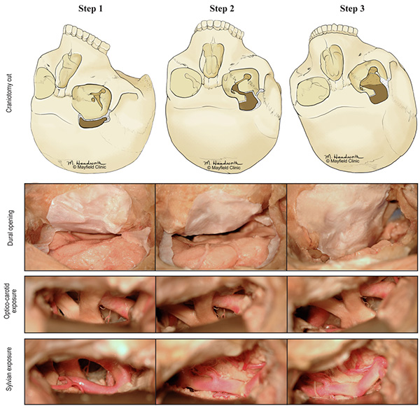
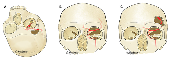
Highlights of the anterolateral progression of surgical exposure for stepwise expansion of the transorbital approach, showing drilling of the lesser (Step 2) and greater sphenoid wings (Step 3) and dural opening in each step. For optico-carotid exposure, notice progressive change from a vertical view of the optic nerve to a more lateral exposure looking across the chiasm. Images on the right highlight sylvian exposure. In example of a high MCA, most of M1’s exposure can be seen in a standard transorbital craniotomy. In other specimens, drilling of the lesser sphenoid wing allows full visualization of the M1 out to the bifurcation. Step 3, or drilling the greater wing nearly to the floor of the middle fossa, provides increased M2 exposure and easier working angles. With permission from Mayfield Clinic.
Successful Bedside Management of a Near-universally Fatal Complication in the NeuroICU: A case report of Tracheal Innominate Artery Fistula following Percutaneous Dilatational Tracheostomy (PDT)
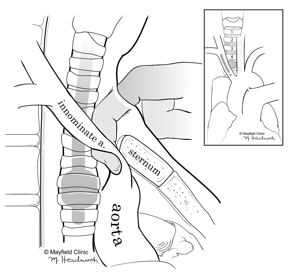
Lateral view illustrating the technique of occluding the innominate artery against the sternum to stop bleeding in case overinflating the tracheal cuff was not enough. [inset] Anterior view demonstrating the close proximity of a low lying tracheostomy (grey circle) below the 4th tracheal ring to the innominate artery. Printed with permission from the Mayfield Clinic.
Illustrations for Supraorbital Versus Superior Eyelid Transorbital (Seto) Approaches: A Qualitative And Quantitative Anatomic Study; manuscript submitted and pending acceptance from Operative Neurosurgery. Alberto Di Somma1, MD, Norberto Andaluz2, MD, Luigi Maria Cavallo1, MD, PhD Jeffrey T. Keller2, PhD, Domenico Solari1, MD, PhD, Lee A. Zimmer3, MD, Matteo de Notaris4, MD, PhD, Mario Zuccarello2, MD, Paolo Cappabianca1, MD
Illustrations showing three successive craiotomies; note the different exposure of the lateral and parasellar compartments of the middle cranial fossa. Supraorbital approach (a); Endoscopic transorbital approach (b); Extended supraorbital approach (transorbital perspective). With permission from Mayfield Clinic.


