

This was a surgical sequence created to explain a method for protecting the brain (cerebroprotection) during heart surgery, in this case the repair of an aortic aneurysm. While it looks complicated - it was done for a cardiac surgeon audience - the important thing to follow is the thick red tube. With the heart stopped, that tube is providing oxygenated blood from the bypass machine to the brain and body. The rest is a tricky set of clamping manoeuvers to maintain as much blood perfusion as possible while isolating the surgical area.
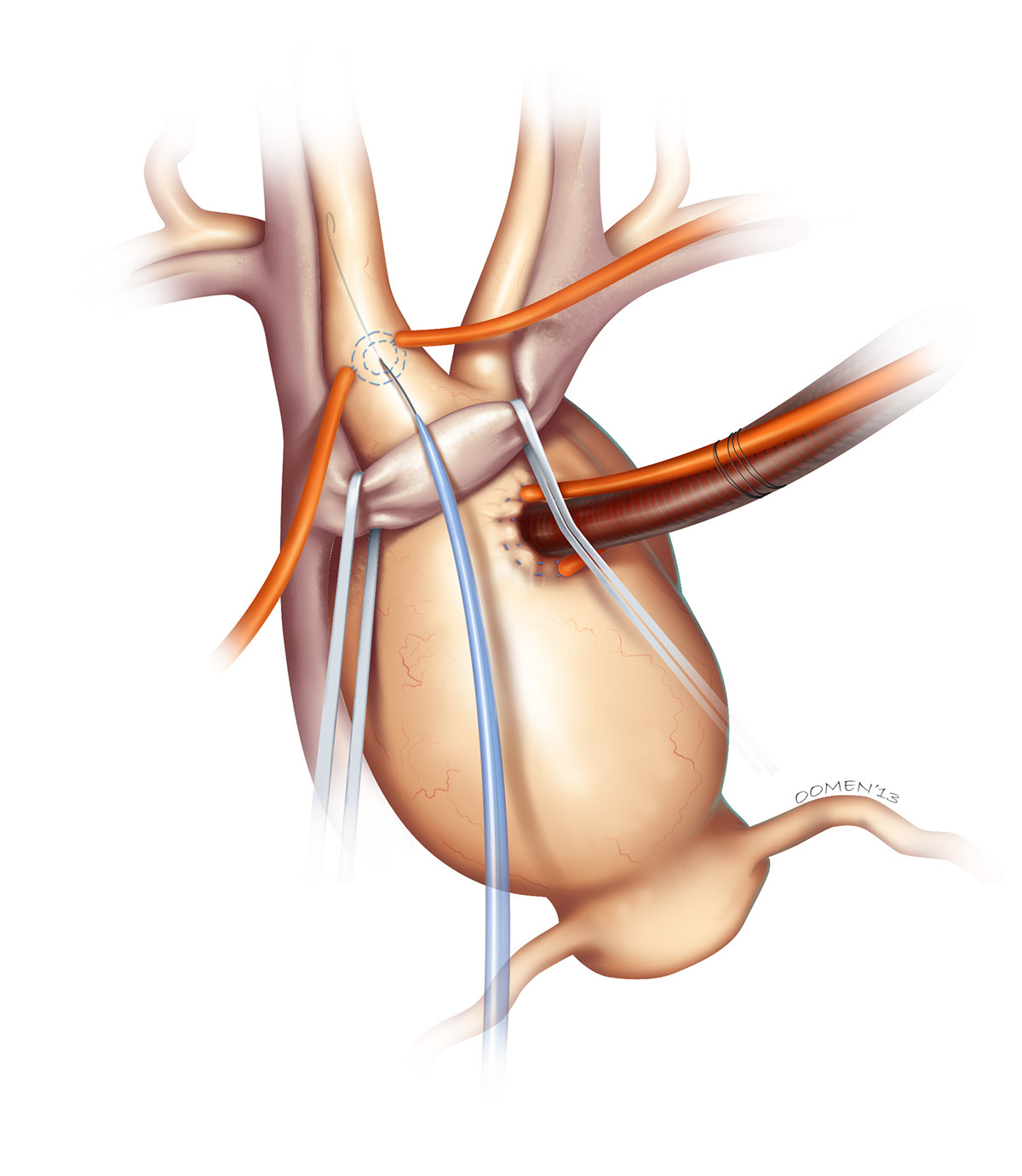
This is the first figure in the sequence, showing the retraction of the inominate vein and canulation of the right brachiocepahlic artery. At this point the aneurysmal aortic arch has also been canulated and put on the bypass machine.

With the right brachiocephalic artery canulated and placed on the bypass machine, the brain is nicely perfused and protected. The aneurysm can be excised and the appropriate graft sewn in place.

Once the top of the graft is sutured snuggly into position, air bubbles are removed and a clamp is placed across the graft below the arm. The bypass line is quickly plugged into the arm of the graft, restoring perfusion to the whole body while the lower end of the graft is sutured onto the aortic root.

The following are some of my (many) sketches done prior to doing any final renderings. This represents less than half of the sketches I would normally do for s surgical sequence like this. It's vital to get just about every detail right, not just how things are shown proceduraly, but what tools are shown, what diameter suture, what clamp, what size tube, what elastics, how things are held etc.
I often put a lot of questions on these sketches as well as point out things I've done in error, then send them to my client for clarification. You can see this above with the "oops" and "not sure..." comments written in.
I often put a lot of questions on these sketches as well as point out things I've done in error, then send them to my client for clarification. You can see this above with the "oops" and "not sure..." comments written in.


